
Development of a High-Sensitivity Triple-Band Nano-Biosensor Utilizing Petahertz Metamaterials for Optimal Absorption in Early-Stage Leukemia Detection
Leukemia starts in the bone marrow and affects normal blood cell production. The cancer progresses in two forms: acute, in which the leukemia cells multiply aggressively, and chronic, in which the cells progress gradually. The prevalence of blood cancers is increasing annually due to genetic and environmental factors. Early detection of cancer greatly improves survival rates. However, bone marrow biopsies, the standard leukemia test, are invasive, time-consuming, and prone to human error.
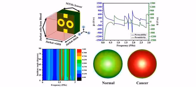
This research presents a new triple-band petahertz nano-biosensor operating at 1.605, 1.848, and 2.424 PHz, achieving a near-perfect absorption (over 98%) with full-angle insensitivity, making it ideal for microwave imaging. The device features a metal-insulator-metal design with a central dipole between two metallic rings of identical diameter.
The rings are made from gold or silver resonators on a silicon dioxide substrate. A solid metallic ground plane prevents all transmission. The design has been improved with a single dipole exhibiting dual peaks near 2.4 PHz. Additional peaks at 1.8 and 2.4 PHz have been achieved by adding dual-ring setups. The final symmetric hybrid creates three sharp, separate absorption peaks using exact geometric symmetry and a 57-micrometer substrate.
Full-wave electromagnetic (EM) simulations use Drude dispersion modeling and fine mesh refinement to accurately capture nanoscale field interactions at the petahertz (PHz) range. Permittivity and permeability analyses show loss peaks that align precisely with the resonance bands. Moreover, the real and imaginary parts of the refractive index and impedance confirm optimal matching in free space, which significantly reduces reflection losses.
Electric fields focus strongly around resonator edges and circular openings at each frequency. Concentration moves from the center at 1.605 PHz to the edges at 2.424 PHz. Surface currents form clockwise rotations concentrated in ring centers, confirming inductive-capacitive coupling for the multiband effect. The biosensor remains polarization-independent, maintaining over 90% absorption efficiency up to 70° of incident angle, making it well-suited for various imaging applications.
The unique EM Signature of Jurkat cells enables precise detection. Researchers model the cells between biological coverslips to mimic the conditions in blood samples closely. Healthy blood has a refractive index of 1.376, while cancerous Jurkat cells have a refractive index of 1.39 due to altered cell membranes, increased metabolic activity, and elevated ionic content.
The higher refractive index alters petahertz wave interactions, causing resonance shifts of 0.00328 PHz in Band 1, 0.02067 PHz in Band 2, and 0.01174 PHz in Band 3. Cancerous samples show stronger red-intensity electric and magnetic fields, providing clear signals to distinguish healthy from cancerous blood. Material choices favor biocompatible gold or silver conductors with stable SiO2 and TiO2 dielectrics. They balance plasmonic efficiency, oxidation resistance, and biomolecular attachment.
PHz operation offers better spatial resolution than terahertz (THz) methods. This nano-biosensor outperforms existing THz and optical sensors through unique PHz triple-band operation, compact design, and precise frequency-shift detection. It enables label-free, real-time monitoring without fluorescent tags or radioactive isotopes. The 100 by 100 nm size supports tight lab-on-chip integration and implantable diagnostics. Future advancements may include multi-cancer biomarker panels, clinical trials, and AI for automated spectral analysis.