



Wireless Peripheral Nerve Recording by Magnetoelectric Nanoparticles
Neuroprostheses and more broadly integrated neurotechnological systems targeting peripheral nerves are fundamental devices for replacing or enhancing motor, sensory, and cognitive functions lost due to neurological diseases or injuries. They are also vital for the medical treatments and monitoring of nerve diseases. The integration of sensors into these systems is crucial for controlling prosthetic limbs and for studying the coding properties of nerve fibers. However, the current sensors' sensitivity-invasiveness trade-off limits the improvements in the performance of bionic devices.
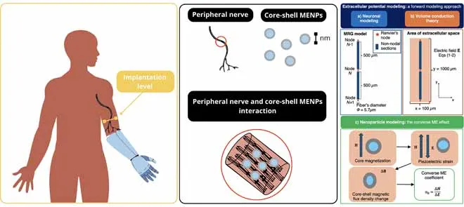
To address this issue, this study proposes using innovative cobalt ferrite (CFO)–barium titanate (BTO) core-shell magnetoelectric nanoparticles (MENPs) as sensors to detect the spontaneous electrical activity of nerve fibers. MENPs are nanometric compounds consisting of a biocompatible piezoelectric shell that coats a magnetostrictive core. Their unique magnetoelectric (ME) effect, characterized by the bidirectional coupling between magnetic and electric fields, arises at the core-shell interface through strain-mediated physical coupling. When an external electric field interacts with these nanoparticles, the magnetization changes. Consequently, even a small electrical signal generated by a nerve cell can cause a measurable change in the particles' magnetic field.
MENPs can be proposed as minimally invasive, highly selective, and wireless sensors due to their biocompatibility, nanometric size, and their ability to change their magnetization in response to changes in the external electric field (converse ME effect). While promising results have been reported on the interaction between MENPs and central nervous system neurons, there is limited information on the interaction with peripheral nerve fibers.
The researchers used a two-stage computational method, i.e., mathematical and computer models, as an alternative to experiments conducted on animals or humans to explore the converse ME effect of MENPs in sensing nerve fibers’ spontaneous electrical activity. The first stage focused on detecting the electrical activity (ranging from approximately -20 to +10 microvolts) of a single peripheral nerve fiber using a double-cable MOTOR (MRG) model, demonstrating that the signals surrounding the nerve are relatively weak. The second stage aimed to analyze the response of CFO-BTO nanoparticles to these weak electric fields. Different factors, including particle sizes, core-to-shell ratios, shapes, magnetic bias fields, and particle arrangements, were studied. Additionally, the physical variables influencing the interaction between MENPs and nerve fibers were quantified.
The results confirmed MENPs' ability to detect the spontaneous electrical activity of nerve fibers and convert it into measurable signals. The maximum local magnetic response reached micro-Tesla levels, thereby significantly amplifying the minimum amplitude measured in human tissues and organs, which is around femto-Tesla. MENPS with a larger core volume and an appropriate core-shell ratio showed higher performance, with a further increase in effectiveness as MENP anisotropy increased. The study also demonstrated the nonlinear influence of an externally applied magnetic bias field on the tuning of MENP response.
The findings of this study offer a valuable strategic framework for adapting the converse ME effect of MENPs to a variety of biomedical applications, paving the way for groundbreaking innovation in integrated neurotechnological systems.