



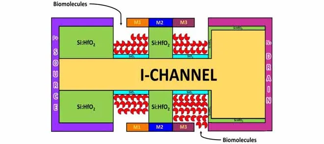
Novel Label-Free Detection of Cancer Biomarkers Using T-Shape Channel and L-Shape Nanocavity in Si:HfO2 Ferroelectric DM-JLTFET With Gate Staggering Biosensor
Early detection of cancer remains the most critical factor in successfully treating the disease and improving patient survival rates. While modern clinical labs can identify specific cancer biomarkers, traditional diagnostic workflows often require complex, time-consuming, and expensive labeling processes. Over the last decade, field-effect transistor (FET) biosensors have emerged as a promising alternative for direct, label-free chemical monitoring. However, standard architectures struggle to consistently isolate neutrally charged biomolecules and suffer severe performance drops when scaled down to nanoscale dimensions.
To address these challenges, the research team engineered a high-performance biosensing device capable of recognizing minute changes with incredible precision. This research introduces a novel architecture called the Ferroelectric Dielectrically Modulated Junctionless Tunnel Field Effect Transistor (FE DM-JLTFET) biosensor. The design is based on two key structural advancements: an intrinsic T-shaped silicon channel and L-shaped nano-cavities that are precisely etched beneath a triple-metal gate.
Instead of traditional oxides, the sensor uses etched cavities in a specialized ferroelectric material known as silicon hafnium dioxide (Si/HfO2). When a blood or tissue sample is applied, target biomarkers naturally accumulate inside these nano-cavities. Since every cell line features a distinct composition of membrane proteins, lipids, and water content, they carry a unique "dielectric footprint." As these molecules fill the cavity, they alter the local electric field. The surrounding ferroelectric layer amplifies this change, dynamically distorting the internal energy bands of the T-shaped channel. This alteration shifts the internal electrical current flowing through the transistor, producing a clear, distinct digital signal without the need for any chemical labels.
Using industry-standard Silvaco TCAD simulation modeling, the device was tested across multiple high-stakes cancer cell lines at specialized terahertz (THz) and gigahertz(GHz) frequencies. The results revealed a highly sensitive capacity to isolate and distinguish between healthy cells and multiple cancer variations:
Kidney Cancer (HEK 293): Differentiated accurately based on distinct electrical shifts at 0.5 THz.
Colorectal Cancer (SW 620): Captured localized cellular differences at 0.5 THz, creating a clear variance in tunneling current.
Osteosarcoma (SaOS-2 and 143B): Successfully classified aggressive bone cancer profiles at 1 GHz based on their high dielectric properties.
This study proved that optimizing the work functions of the triple-metal gate, specifically using a staggered configuration of 3.8/4.6/4.0 eV, significantly increases electron injection, thereby boosting overall sensitivity. The researchers also demonstrated that higher filling levels within the nano-cavities yield stronger signal variations, confirming peak operating efficiency when the cavity is fully utilized.
This compact biosensing device operates efficiently at a 2.4 GHz frequency with minimal power consumption. At just 80 nanometers, the sensor effectively overcomes the major short-channel electrical leakage that plagues conventional electronics. Its fabrication process leverages standard semiconductor manufacturing techniques, making the device highly scalable and cost-effective. This technology lays the foundation for a rapid, ultra-sensitive, point-of-care screening tool that could bring routine, non-invasive early cancer diagnostics directly to a patient’s bedside.